
vision for the future
Ramole Eye Hospital in Nashik is NABH Accredited Advanced Eye Care Centre in Nashik with all facilities like Advance PHACO Micro Surgery (MICS), Toric Lens Implant, New Refractive Tricofocal Implant, Contoura Lasik Vision Surgery, Advanced Surface Treatment, Phakic Implant (Phakic Lens/ICL), Keratoconus Treatment (C3R) etc. with Specialised & Customized Facilities available for All Age Groups including Pediatric to Senior Citizens. We provide all types of Mediclaim, ESIC, Facilities too.
CATARACT SURGERY HELPS IN FOLLOWING EYE PROBLEMS

Nearsightedness (Myopia)
Myopia, commonly known as nearsightedness, is a refractive error of the eye where close objects are seen clearly, but distant objects appear blurry.
Farsightedness (Hyperopia)
Farsightedness, also known as hyperopia, is a common refractive error in the eye where distant objects can be seen more clearly than nearby ones.
Astigmatism (Blurry Vision)
Astigmatism is a common refractive error in the eye that occurs when the cornea or lens has an irregular shape, causing blurred or distorted vision.
Cataract PHACO surgery
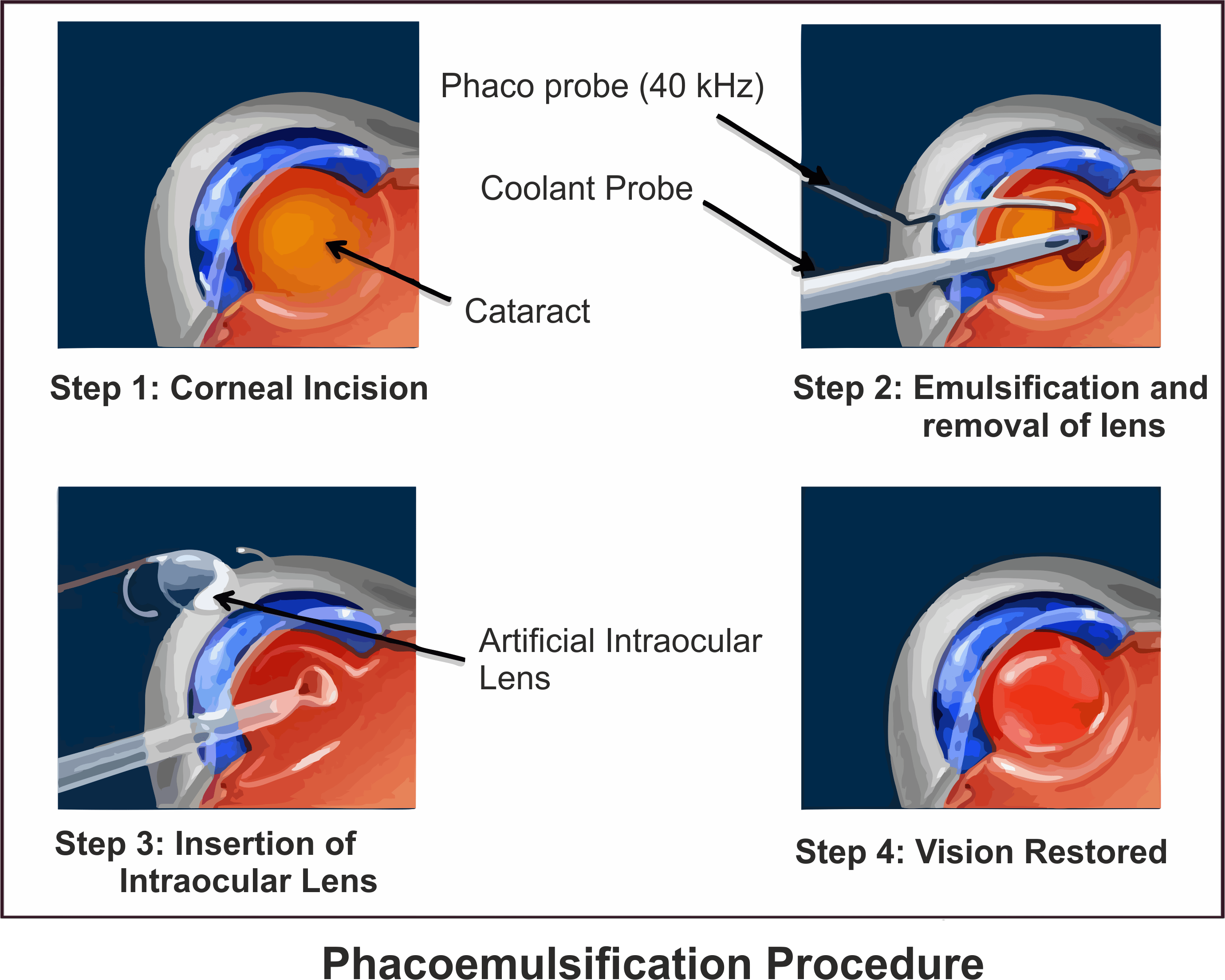
Cataract surgery has evolved significantly over the years, and one of the ground-breaking techniques that have revolutionized the field is Phacoemulsification, commonly known as PHACO. This advanced surgical procedure has transformed cataract removal into a minimally invasive and highly effective process, with the added benefit of a tiny 2.8mm incision. Let’s delve into the marvel of PHACO and explore how it has become the gold standard in cataract surgery.
Here’s an overview of the PHACO (Phacoemulsification) procedure:
1) Anesthesia:
The process begins with the administration of local anesthesia to numb the eye, ensuring the patient’s comfort during the procedure.
2) Incision Creation:
A small incision, typically less than 3 millimeters, is made at the edge of the cornea to access the eye’s interior.
3) Capsulotomy:
A circular opening is created in the eye’s natural lens capsule, allowing access to the cataract-affected lens.
4) Phacoemulsification:
Ultrasonic vibrations are used to break the cloudy lens (cataract) into tiny fragments, which are then gently suctioned out through the probe.
5) Lens Implantation:
Once the cataract is removed, an artificial intraocular lens (IOL) is inserted through the same small incision to replace the natural lens.
6) Wound Closure:
In most cases, the self-sealing incision doesn’t require stitches. It naturally closes and begins healing.
7) Posterior Capsule Polishing:
If needed, the surgeon may perform polishing of the posterior capsule to ensure clarity and prevent future clouding.
8) IOL Adjustment:
The surgeon ensures the proper positioning and adjustment of the artificial lens for optimal vision correction.
9) Final Inspection:
The surgeon verifies the integrity of the incision and the stability of the intraocular lens.
10) Post-Operative Care:
Patients receive instructions for post-operative care, including the use of prescribed eye drops to prevent infection and aid in healing.
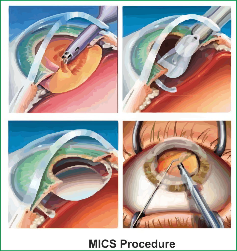
Micro Incision Cataract Surgery (MICS) Surgery
Micro Incision Cataract Surgery (MICS) is a state-of-the-art surgical technique that aims to remove cataracts with unparalleled precision and minimal invasiveness. Unlike traditional cataract surgery, which involves larger incisions, MICS utilizes an incredibly small 1.2mm incision, significantly reducing the surgical trauma to the eye.
Here’s an overview of the MICS (Micro Incision Cataract Surgery) procedure:
1) Anesthesia:
Local anesthesia is administered to numb the eye, ensuring patient comfort during the surgery.
2) Incision Creation:
A tiny incision, typically around 1.8 millimeters or smaller, is made at the edge of the cornea.
3) Capsulotomy:
A circular opening is created in the lens capsule to access the cataract.
4) Phacoemulsification:
Ultrasonic energy is used to break the cataract into small pieces, which are then suctioned out through the micro-incision.
5) Lens Insertion:
An artificial intraocular lens (IOL) is folded and inserted through the micro-incision. Once inside the eye, it unfolds to replace the natural lens.
6) Wound Closure:
The self-sealing incision typically does not require sutures and begins to heal naturally.
7) IOL Positioning:
The surgeon ensures proper placement and alignment of the intraocular lens for optimal vision correction.
8) Posterior Capsule Polishing:
If necessary, the surgeon may perform polishing of the posterior lens capsule for clarity.
9) Final Inspection:
The surgeon checks the incision and confirms the stability of the intraocular lens.
10) Post-Operative Care:
Patients receive instructions for post-operative care, including the use of prescribed eye drops to prevent infection and aid in the healing process.