
Glaucoma Disease
get in touch with us
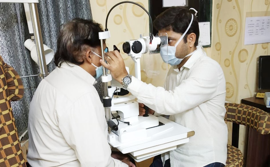
Manage glaucoma with confidence at Ramole Eye Hospital. Our specialized team offers expert care and advanced treatments to effectively manage this condition. Trust us for personalized care and support on your journey to preserving vision.
frequently
asked questions
Explore the essential facts surrounding Glaucoma and gain insights into effective management strategies.
Glaucoma is a disease of the optic nerve – the part of the eye that carries the images we see to the brain. The optic nerve is made up of many nerve fibers, like an electric cable containing numerous wires. When damage to the optic nerve fibers occurs, blind spots develop. These blind spots usually go undetected until the optic nerve is significantly damaged. If the entire nerve is destroyed, blindness results.
Early detection and treatment by your ophthalmologist (Eye M.D.) are the keys to preventing optic nerve damage and blindness from glaucoma. Glaucoma is a leading cause of blindness in the United States, especially for older people. but loss of sight from glaucoma can often be prevented with early treatment.
Chronic-open-angle glaucoma: This is the most common form of glaucoma in the United States. The risk of developing chronic open-angle glaucoma increases with age. The drainage angle of the eye becomes less efficient over time, and pressure within the eye gradually increases, which can damage the optic nerve. In some patients, the optic nerve becomes sensitive even to normal eye pressure and is at risk for damage. Treatment is necessary to prevent further vision loss. See a short video on Open Angle Glaucoma.
Typically, open-angle glaucoma has no symptoms in its early stages, and vision remains normal. As the optic nerve becomes more damaged, blank spots begin to appear in your field of vision. You typically won’t notice these blank spots in your day-to-day activities until the optic nerve is significantly damaged and these spots become large. If all the optic nerve fibers die, blindness results. Follow this link to a short video on Glaucoma and Blind Spots.
Closed-angle glaucoma: Some eyes are formed with the iris (the colored part of the eye) too close to the drainage angle. In these eyes, which are often small and farsighted, the iris can be sucked into the drainage angle and block it completely. Since the fluid cannot exit the eye, pressure inside the eye builds rapidly and causes an acute closed-angle attack.
Clear liquid called aqueous humor circulates inside the front portion of the eye. To maintain a healthy level of pressure within the eye, a small amount of this fluid is produced constantly while an equal amount flows out of the eye through a microscopic drainage system. (This liquid is not part of the tears on the outer surface of the eye.)
If the drainage angle is blocked, excess fluid cannot flow out of the eye, causing the fluid pressure to increase.
Because the eye is a closed structure, if the drainage area for the aqueous humor-called the drainage angle-is blocked, the excess fluid cannot flow out of the eye. Fluid pressure within the eye increases, pushing against the optic nerve and causing damage.
Symptoms of Glaucoma May Include:
Blurred vision
Severe eye pain
Headache
Rainbow-colored halos around lights
Nausea and vomiting
This is a true eye emergency. If you have any of these symptoms, call your ophthalmologist immediately. Unless this type of glaucoma is treated quickly, blindness can result. Unfortunately, two-thirds of those with closed-angle glaucoma develop it slowly without any symptoms prior to an attack.
The most important risk factors include:
- Age
- Elevated eye pressure
- Family history of glaucoma
- Farsightedness or nearsightedness
- Past eye injuries
- Thinner central corneal thickness
- Systemic health problems, including diabetes, migraine headaches, and poor circulation.
- Measure your intraocular pressure (tonometry)
- Inspect the drainage angle of your eye (gonioscopy)
- Evaluate whether or not there is any optic nerve damage (ophthalmoscopy)
- Test the peripheral vision of each eye (visual field testing, or perimetry)
- Pachymetry is the test used to measure corneal thickness. (pachymetry)
- Optical coherence tomography (OCT) is a type of non-invasive imaging test used to observe each distinctive layer of the retina. This test may be recommended.
- Visual field testing is used to monitor the development of blind spots and peripheral or side vision.
Some of these tests may not be necessary for everyone. These tests may need to be repeated on a regular basis to monitor any changes in your condition.
Loss of vision can be prevented Regular medical eye exams can help prevent unnecessary vision loss.
Book An Appointment Today.