Retinal Diseases

Retinal diseases affect the sensitive tissue at the back of the eye responsible for capturing light and sending signals to the brain.

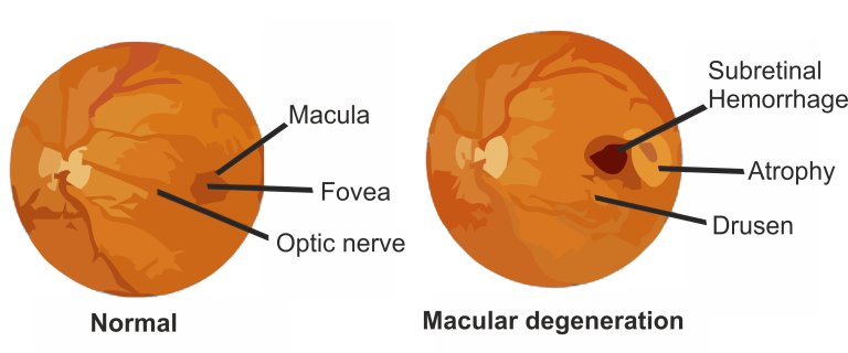
Age-Related Macular Degeneration (AMD) is a progressive eye disease affecting the macula, the central portion of the retina responsible for sharp, central vision. It is the leading cause of severe vision loss in people over 50.

The primary cause of AMD is aging, particularly in individuals over 50. Other risk factors include a family history of AMD, smoking, hypertension, and certain genetic factors.

1. Blurred or distorted central vision.
2. Difficulty recognizing faces.
3. Straight lines appear wavy.

Dry AMD (Non-Neovascular): Characterized by the gradual breakdown of light-sensitive cells in the macula.
Wet AMD (Neovascular): Involves the growth of abnormal blood vessels beneath the retina, leading to leakage of blood and fluid.

Dilated Eye Exam: A comprehensive eye examination includes examining the retina for signs of AMD.
Amsler Grid Test: A grid with straight lines is used to detect any distortion in the central vision.
Optical Coherence Tomography (OCT): Produces detailed cross-sectional images of the retina.

Diabetic Retinopathy is a diabetes-related eye disease that affects the retina. It arises due to damage to the blood vessels in the light-sensitive tissue at the back of the eye (retina) and is a leading cause of vision loss among individuals with diabetes.

The primary cause of Diabetic Retinopathy is prolonged high blood sugar levels associated with diabetes. Over time, these elevated levels can damage the tiny blood vessels that nourish the retina.

1. Blurred or fluctuating vision.
2. Impaired color vision.
3. Floaters or dark spots.
4. Difficulty in recognizing faces.

Non-Proliferative Diabetic Retinopathy (NPDR): Characterized by weakened blood vessels that may leak, leading to swelling of the macula.
Proliferative Diabetic Retinopathy (PDR): Involves the growth of abnormal blood vessels, which can bleed into the vitreous, causing vision loss.

Dilated Eye Exam: Essential for detecting changes in the retina.
Optical Coherence Tomography (OCT): Provides detailed cross-sectional images of the retina.

Hypertensive Retinopathy is a condition characterized by damage to the retina’s blood vessels due to chronic high blood pressure. Prolonged hypertension can lead to changes in the retina, affecting vision.

Consistently elevated blood pressure puts strain on delicate retinal blood vessels, causing them to narrow, leak, or become blocked. This can lead to various retinal changes associated with Hypertensive Retinopathy.

Visual Impairment: Blurred or distorted vision.
Headaches: Especially in cases of severe hypertensive retinopathy.
Reduced Visual Acuity: Difficulty in seeing clearly.

  1. Grade 1 – Mild Hypertensive Retinopathy: Early signs of hypertensive retinopathy, including mild narrowing of the retinal arteries (arteriolar narrowing) and a slightly silver or copper-wire appearance.

  2. Grade 2 – Moderate Hypertensive Retinopathy: Progression to more pronounced arteriolar narrowing, along with additional signs such as arteriovenous nicking (compression of veins at arteriovenous crossings) and retinal hemorrhages.

  3. Grade 3 – Severe Hypertensive Retinopathy: Significant arteriolar narrowing, arteriovenous nicking, and more widespread retinal hemorrhages. Cotton-wool spots (soft exudates) may also be present, indicating areas of retinal nerve fiber layer infarction.

  4. Grade 4 – Malignant (Accelerated) Hypertensive Retinopathy: This is a severe and urgent form of hypertensive retinopathy. It involves all the signs of Grade 3 retinopathy but with the addition of optic nerve swelling (papilledema). Malignant hypertensive retinopathy requires immediate medical attention.

Funduscopy: Examining the retina to identify characteristic signs like narrowing of arteries, flame-shaped hemorrhages, and cotton-wool spots.
Optical Coherence Tomography (OCT): Imaging to assess retinal thickness and integrity.

Retinal Detachment is a serious eye condition where the retina, the thin layer of tissue at the back of the eye responsible for vision, pulls away from its normal position. This detachment can lead to severe visual impairment if not promptly addressed.

Tears or Breaks: Breaks or tears in the retina allow fluid to accumulate, causing detachment.
Age and Nearsightedness: More common in older adults and those with extreme nearsightedness.
Trauma or Injury: Direct trauma to the eye can cause detachment.
Family History: There might be a genetic predisposition.

Sudden Floaters: Seeing floating specks or cobweb-like floaters in the field of vision.
Flashes of Light: Perceiving sudden flashes of light.
Shadow or Curtain Effect: A sensation of a shadow or curtain descending over the visual field.

Rhegmatogenous: Caused by a hole or tear in the retina.
Tractional: Resulting from scar tissue pulling the retina.
Exudative: Fluid buildup behind the retina without a tear.

Retinal Examination: By dilating the pupil, an ophthalmologist can examine the retina for detachment signs.
Ultrasound: Useful in identifying detachment in specific cases.
Ophthalmoscopy: Examination of the retina.

Vitritis is an inflammatory condition characterized by inflammation of the vitreous humor, the gel-like substance filling the back part of the eye.

Infections: Viral, bacterial, fungal, or parasitic.
Autoimmune Diseases: Conditions like uveitis.
Inflammatory Disorders: Such as sarcoidosis or Behçet’s disease.
Posterior Uveitis: Inflammation affecting the back of the eye.

Blurred Vision: Impaired visual clarity.
Floaters: Dark spots or lines moving across vision.
Eye Pain: Discomfort or pain may be present.
Photophobia: Sensitivity to light.

  1. Infectious Vitritis: Caused by infections such as viral, bacterial, or fungal agents. These infections can trigger inflammation in the vitreous.

  2. Non-Infectious Vitritis: Inflammation not related to an infection. This type can be associated with autoimmune diseases, inflammatory conditions, or unknown factors.

  3. Posterior Vitreous Detachment (PVD): As the vitreous humor naturally separates from the retina with age, it can lead to vitritis-like symptoms, including floaters and light flashes.

  4. Intermediate Uveitis: Also known as pars planitis, this is a type of uveitis that involves inflammation in the middle portion of the eye, including the vitreous.

  5. Toxoplasmosis-Related Vitritis: Vitritis can occur as a result of toxoplasmosis, an infection caused by the parasite Toxoplasma gondii.

Ophthalmic Examination: Comprehensive eye examination.
Optical Coherence Tomography (OCT): Imaging for detailed retina evaluation.
Blood Tests: To identify underlying infections or autoimmune conditions.

Vitreous Hemorrhage refers to the presence of blood within the vitreous humor, the gel-like substance that fills the back part of the eye.

Diabetic Retinopathy: Abnormal blood vessels in the retina.
Retinal Tears or Detachment: Trauma or age-related conditions.
Macular Degeneration: Breakdown of macula blood vessels.
Eye Injuries: Direct trauma to the eye.
Blood Vessel Abnormalities: Conditions like retinal vein occlusion.

Sudden Floaters: Dark spots or lines moving across vision.
Blurred or Cloudy Vision: Impaired visual clarity.
Flashes of Light: Occasional flashes in the affected eye.

1. Traumatic Vitreous Hemorrhage:
Hemorrhage occurs due to physical damage, such as a direct blow to the eye.
2. Age-Related Vitreous Hemorrhage:
Commonly seen in elderly individuals as the vitreous humor undergoes changes, leading to bleeding.
3. Diabetic Vitreous Hemorrhage:
Elevated blood sugar levels can damage blood vessels in the retina, leading to bleeding into the vitreous.
4. Retinal Tear or Detachment-Related Vitreous Hemorrhage:
Blood from retinal vessels may leak into the vitreous, especially in cases of retinal tears or detachments.
5. Vascular Abnormality-Related Vitreous Hemorrhage:
Unusual vascular structures can lead to bleeding into the vitreous.
6. Hypertensive Vitreous Hemorrhage:
Elevated blood pressure can damage blood vessels in the eye, resulting in bleeding.
7. Blood Disorders-Related Vitreous Hemorrhage:
Disorders like hemophilia or sickle cell anemia can contribute to bleeding in the vitreous.

Ocular Ultrasound: Imaging technique to visualize the vitreous and retina.
Fluorescein Angiography: Tracing blood flow in the retina.

A Macular Hole is a small break or hole in the macula, the central part of the retina responsible for sharp, central vision. This condition may result in vision distortion or blurriness.

Age: More prevalent in individuals over 60.
Vitreous Detachment: When the vitreous, the gel-like substance in the eye, pulls away from the macula, it can cause a hole.
Eye Trauma: Severe injury to the eye can contribute.

Blurred or Distorted Central Vision: Straight lines may appear wavy or distorted.
Decreased Central Vision: A gradual loss of detailed vision.
Visual Distortions: Objects may appear smaller or farther away.

Foveal Dehiscence: An early stage with minimal symptoms.
Partial-Thickness Hole: A partial break in the macula.
Full-Thickness Hole: A complete hole from the inner to outer retina.

Optical Coherence Tomography (OCT): Imaging to visualize the macula’s cross-section.
Fluorescein Angiography: Injecting dye to highlight blood vessels and assess the extent of damage.

Central Retinal Artery Occlusion (CRAO) is a vascular emergency characterized by the sudden blockage of the central retinal artery, leading to a rapid and severe decrease in blood supply to the retina.

Atherosclerosis: Build-up of plaque in the arteries.
Cardiovascular Diseases: Hypertension, heart valve disorders.
Blood Clot Disorders: Conditions affecting blood clotting.

Sudden, Profound Vision Loss: Typically in one eye.
Central Scotoma: Dark spot at the center of vision.
Retinal Pallor: Whitening of the retina upon examination.

Non-Arteritic CRAO: More common, often associated with atherosclerosis.
Arteritic CRAO: Linked to inflammation of the arteries, particularly seen in giant cell arteritis.

Fluorescein Angiography: Visualizing blood flow in the retina.
Ocular Coherence Tomography (OCT): Assessing retinal thickness and structural changes.

Central Retinal Vein Occlusion (CRVO) is a vascular disorder affecting the retina, where the central vein that drains blood from the retina becomes blocked, leading to impaired blood flow and potential vision loss.

Atherosclerosis: Hardening and narrowing of the arteries.
Glaucoma: Increased pressure in the eye.
Diabetes: Elevated blood sugar levels.

Sudden Blurred Vision: Usually affecting one eye.
Floaters: Dark spots or lines in the visual field.
Visual Field Loss: Especially in advanced cases.

Ischemic CRVO: Severe blockage leading to inadequate blood supply, posing a higher risk of vision loss.
Non-Ischemic CRVO: Partial blockage with better-preserved blood flow and a relatively lower risk of severe vision impairment.

Fluorescein Angiography: Dye-based imaging to evaluate blood flow in the retina.
Optical Coherence Tomography (OCT): Assessing retinal thickness and identifying fluid accumulation.

Central Serous Retinopathy (CSR) is a retinal disorder characterized by the accumulation of fluid beneath the central macula, leading to distorted vision.

Stress: Chronic stress may contribute.
Corticosteroid Use: Systemic or local steroid use.
Hypertension: High blood pressure.
Male Predominance: More common in males.

Blurred or Distorted Vision: Central vision impairment.
Diminished Color Perception: Colors may appear less vibrant.
Metamorphopsia: Visual distortion of straight lines.

  1. Acute Central Serous Retinopathy: The most common form, characterized by a sudden onset of visual disturbances. It is often associated with the accumulation of fluid under the central macula.

  2. Chronic Central Serous Retinopathy: In some cases, the condition may become chronic, with persistent or recurrent episodes of fluid leakage. Chronic CSR can lead to long-term visual changes and may require ongoing management.

  3. Multifocal Central Serous Retinopathy: In this type, multiple areas of fluid accumulation occur across the retina, leading to a more widespread and variable pattern of visual symptoms.

  4. Bullous Central Serous Retinopathy:Bullous CSR is characterized by the formation of large, dome-shaped elevations of the retina due to the accumulation of subretinal fluid. This type may have a more severe impact on visual function.

  5. Recurrent Central Serous Retinopathy: Some individuals experience recurrent episodes of CSR, with periods of fluid leakage followed by resolution. Recurrent CSR may require close monitoring and tailored management strategies.

Ophthalmic Examination: Dilated eye exam to assess the retina.
Fluorescein Angiography: Dye injection to visualize blood flow in the retina.
Optical Coherence Tomography (OCT): Imaging for detailed retinal assessment.

Scroll to Top