Common Eye Problems

myopia

Our eyes play a vital role in our daily lives, allowing us to perceive the world around us. However, various factors can impact our vision and eye health, leading to common eye problems. Understanding these common eye problems and their symptoms is crucial for seeking timely care and maintaining optimal eye health. Regular eye check-ups play a key role in detecting issues early, allowing for effective management, and preserving the gift of sight.

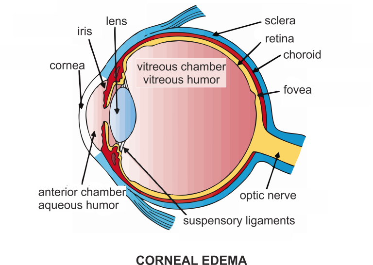
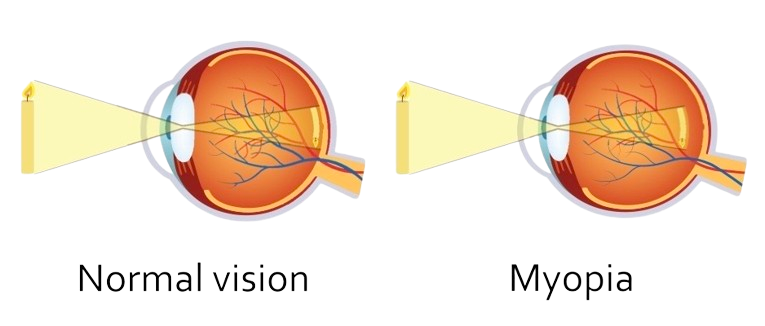
Myopia, commonly known as nearsightedness, is a refractive error of the eye where close objects are seen clearly but distant objects appear blurry. This occurs when the eyeball is too long or the cornea has too much curvature, causing light entering the eye to focus in front of the retina instead of directly on it.

1. Genetics: Myopia often runs in families, suggesting a strong genetic component.
2. Environmental Factors: Prolonged proximity to work, such as reading or computer use, can contribute.
3. Eye Strain: excessive strain on the eyes, especially during activities requiring close focus.

1. Blurred vision for distant objects.
2. Eye strain or discomfort.
3. Squinting to see clearly.

Low Myopia:
Diopter Range: Up to -3.00 D
Individuals with mild myopia may experience slight blurriness for distant objects but can usually see well without corrective lenses.

Moderate Myopia:
Diopter Range: Between -3.00 D and -6.00 D
Blurred vision for distant objects becomes more noticeable. Corrective lenses are usually prescribed.

High Myopia:
Diopter Range: Beyond -6.00 D
Severe blurriness for distant objects. There is a higher risk of associated eye conditions such as retinal detachment, glaucoma, and cataracts.

Apart from the severity classification, myopia can also be categorized based on its progression:

Simple Myopia:
Stabilizes in adulthood without significant progression.

Pathological Myopia:
Associated with structural changes in the eye, increasing the risk of complications. This includes a higher risk of retinal detachment, glaucoma, and macular degeneration.
It’s important to note that myopia can also be categorized by the age of onset:

Congenital Myopia:
Present at birth or develops in infancy.

Acquired Myopia:
Develops later in childhood or adolescence.

1. Prescription Glasses or Contact Lenses: Corrective lenses help focus light directly on the retina.
2. Refractive Surgery: Procedures like LASIK reshape the cornea for clearer vision.
3. Orthokeratology: the use of special contact lenses to reshape the cornea temporarily.

Hyperopia

hyperopia

Farsightedness, also known as hyperopia, is a common refractive error in the eye where distant objects can be seen more clearly than nearby ones. In a farsighted eye, light entering the eye focuses behind the retina instead of directly on it.

1. Light Focusing Behind the Retina: In a farsighted eye, the eyeball is too short or the cornea has too little curvature. As a result, light entering the eye is not focused on the retina but converges behind it.
2. Blurred Near Vision: Farsighted individuals may experience difficulty seeing objects up close, such as when reading or doing close-up tasks. The ability to see distant objects is generally better.
3. Common Vision Issue: Hyperopia can be present from birth and may change with age. It often coexists with presbyopia, an age-related condition that affects near vision.

1. Blurred Near Vision: Farsighted individuals often experience difficulty seeing objects up close, such as when reading, working on a computer, or doing close-up tasks.
2. Eye Strain: Trying to focus on close objects can lead to eye strain, discomfort, and fatigue.
3. Headaches: Extended periods of close work may result in headaches, particularly around the forehead and temples.
4. Difficulty Seeing Up Close: People with hyperopia may find it challenging to perform tasks that require clear vision at close distances.
5. Eye Discomfort: Farsightedness can cause general eye discomfort or a feeling of tension, especially during near tasks.
6. Squinting: Farsighted individuals may unconsciously squint to help bring close-up objects into focus, which can alleviate the blurriness temporarily.

1. Diagnosis: An eye exam, including a refraction test, helps determine the degree of farsightedness. The prescription is measured in positive diopters.
2. Vision Correction: Eyeglasses or contact lenses with a prescription that converges light onto the retina can correct farsightedness. In some cases, refractive surgery, such as LASIK, may be an option.
3. Regular Eye Check-ups: Monitoring vision changes and adjusting corrective prescriptions are important to maintain optimal vision health.
Farsightedness is a common vision issue that can be effectively corrected with appropriate lenses or surgical interventions. Regular eye check-ups are essential for managing vision changes over time.

1. Prescription Glasses or Contact Lenses: Corrective lenses help focus light directly on the retina.
2. Refractive Surgery: Procedures like LASIK reshape the cornea for clearer vision.
3. Orthokeratology: the use of special contact lenses to reshape the cornea temporarily.

Astigmatism

astigmatism

Astigmatism is a common refractive error in the eye that occurs when the cornea or lens has an irregular shape, causing blurred or distorted vision. In a healthy eye, the cornea and lens are ideally shaped like a round ball. However, in astigmatism, these structures are more oval or football-shaped.

  1. Genetic factors
  2. Changes in the eye’s tissues over time
  3. Eye injuries.
  1. Blurred Vision: Astigmatism can lead to distorted or blurred vision at various distances.
  2. Eye Strain: Individuals with astigmatism may experience eye strain, discomfort, or headaches, especially after extended periods of close-up work.
  3. Squinting: Squinting may occur in an attempt to see more clearly.
  1. Regular Astigmatism: The cornea or lens has a consistent curvature in one principal meridian, causing clear vision along one axis.
  2. Irregular Astigmatism: The curvature varies in different meridians, often resulting from corneal scarring or certain eye conditions.
  1. Eyeglasses: Prescription glasses with cylindrical lenses can compensate for the uneven curvature.
  2. Contact Lenses: Toric contact lenses are designed to correct astigmatism, offering an alternative to glasses.
  3. Refractive Surgery: LASIK or other refractive surgeries can reshape the cornea, correcting astigmatism permanently.

Squint (Strabismus)

squint

Squint, or strabismus, is a visual condition where the eyes are misaligned and do not look in the same direction. One eye may turn inward, outward, upward, or downward, while the other remains straight.

1. Muscular Imbalance: Weakened or imbalanced eye muscles.
2. Nerve Abnormalities: Issues with the nerves controlling eye muscles.
3. Refractive Errors: Uncorrected farsightedness, nearsightedness, or astigmatism.
4. Genetic Factors: Family history of strabismus increases the risk.
5. Eye Injuries or Diseases: Trauma or certain eye conditions can contribute.

1. Misalignment: Eyes not pointing in the same direction.
2. Double Vision: Overlapping or blurry vision.
3. Eye Fatigue: Strain or tiredness in the eyes.
4. Head Tilting: Tilting the head to align the eyes.
5. Poor Depth Perception: Difficulty judging distances.

1. Esotropia: Inward turning of one or both eyes.
2. Exotropia: Outward turning of one or both eyes.
3. Hypertropia: Upward turning of one eye.
4. Hypotropia: Downward turning of one eye.

  1. Visual Acuity Test:

    • Measures the clarity of vision in each eye using an eye chart.
  2. Cover Test:

    • Detects misalignment by covering one eye at a time, observing the movement of the covered eye when the other is focused on an object.
  3. Corneal Light Reflex Test (Hirschberg Test):

    • Checks the alignment of the eyes by observing the reflection of light on the corneas.
  4. Prism and Cover Test:

    • Uses prisms to measure the amount and direction of eye misalignment.
  5. Refraction Test:

    • Determines the need for glasses or contact lenses and helps assess the degree of refractive error.
  6. Ocular Motility Test:

    • Evaluates eye movements in different directions, identifying limitations or abnormalities.
  7. Stereopsis Test:

    • Assesses depth perception by measuring the ability of both eyes to work together.
  8. Specialized Eye Imaging:

    • In some cases, imaging techniques like MRI or CT scans may be used to assess the eye muscles and surrounding structures.

Corneal Abrasion

Scratching or injury to the corneal surface is called as Corneal Abrasion. 

Foreign Objects: Particles like dust, sand, or metal entering the eye.
Contact Lenses: Incorrect use, poor hygiene, or overuse.
Scratched Cornea: Rubbing the eye vigorously.
Chemical Exposure: Contact with harmful substances.
Blunt Trauma: Injury, accidents, or sports-related incidents.

Eye Pain: Intense discomfort and pain.
Watery Eyes: Excessive tearing.
Light Sensitivity: Photophobia.
Blurry Vision: Impaired or fluctuating vision.
Foreign Body Sensation: Feeling like something is in the eye.
Redness: Bloodshot appearance.

Superficial Abrasion: Limited to the outermost corneal layer.
Corneal Erosion: Repeated breakdown of corneal epithelium.
Recurrent Erosion Syndrome: Chronic corneal erosions.

  1. Slit-lamp Examination: This specialized microscope allows the eye care professional to examine the cornea and other structures of the eye in detail.

  2. Fluorescein Staining: A special dye, usually fluorescein, is applied to the eye. The dye adheres to damaged areas of the cornea, making abrasions more visible under certain lighting conditions.

  3. Visual Acuity Test: This common eye test measures how well you can see at various distances. It helps determine the extent of vision impairment caused by the abrasion.

  4. Tonometry: This test measures the pressure inside the eye, which can help rule out conditions like glaucoma.

  5. Pupil Dilation: Dilating the pupils with special eye drops allows for a more comprehensive examination of the eye’s internal structures.

Corneal Edema

Corneal Edema is a condition characterized by the accumulation of fluid in the cornea, the transparent front part of the eye.

Corneal Endothelial Dysfunction: A decrease in the function of endothelial cells that maintain corneal clarity.
Corneal Injury or Trauma: Physical damage to the cornea, leading to fluid retention.
Eye Surgery: Certain eye surgeries, like cataract surgery, may contribute to corneal edema.

Blurred Vision: Reduced clarity of vision.
Halos Around Lights: Glare or halos around lights, especially at night.
Eye Discomfort: A feeling of fullness or discomfort in the eye.
Decreased Visual Acuity: Gradual loss of visual sharpness.

Corneal edema may be categorized based on its underlying causes, such as Fuchs’ dystrophy or postsurgical edema.

1.Slit-lamp Examination: This specialized microscope allows for a detailed examination of the cornea and other structures of the eye. It helps the eye care professional visualize any swelling or cloudiness in the cornea.

2. Visual Acuity Test: This common eye test measures how well you can see at various distances. It helps determine the extent of vision impairment caused by corneal edema.

3. Pachymetry: Pachymetry is a diagnostic procedure used to gauge the thickness of the cornea. Increased thickness may indicate the presence of corneal edema.

4. Specular Microscopy: This imaging technique provides a detailed view of the corneal endothelium, the innermost layer of the cornea. Changes in endothelial cells may be observed in cases of corneal edema.

5. Tonometry: This test measures the pressure inside the eye, which can help rule out conditions like glaucoma that may contribute to corneal edema.

6. Corneal Topography: This mapping of the cornea’s surface can reveal irregularities and changes in shape associated with corneal edema.

Scroll to Top