
vision for the future
Ramole Eye Hospital in Nashik is NABH Accredited Advanced Eye Care Centre in Nashik with all facilities like Advance PHACO Micro Surgery (MICS), Toric Lens Implant, New Refractive Tricofocal Implant, Contoura Lasik Vision Surgery, Advanced Surface Treatment, Phakic Implant (Phakic Lens/ICL), Keratoconus Treatment (C3R) etc. with Specialised & Customized Facilities available for All Age Groups including Pediatric to Senior Citizens. We provide all types of Mediclaim, ESIC, Facilities too.
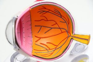
RETINA SURGERY HELPS IN FOLLOWING EYE DISEASES
AMD
Age-Related Macular Degeneration (AMD) is a progressive eye disease affecting the macula responsible for sharp vision.
retinal Detachment
Retinal Detachment is a serious eye condition where the retina, responsible for vision, pulls away from its normal position.
Vitritis
Vitritis is an inflammation of the vitreous humor. It can be diagnosed with Optical Coherence Tomography (OCT).
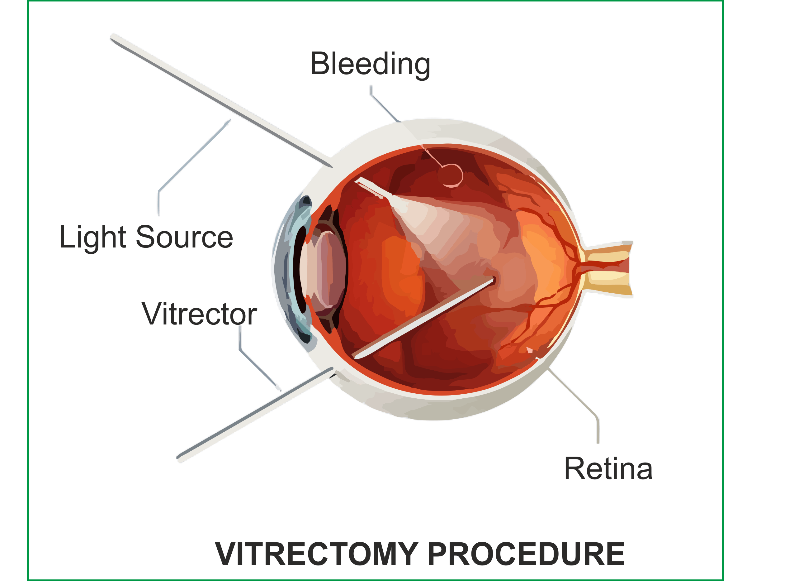
Vitrectomy surgery
A vitrectomy is a surgical procedure performed to remove the vitreous gel from the middle of the eye. This procedure is typically done to treat various eye conditions, including retinal detachment, vitreous hemorrhage, macular hole, epiretinal membrane, diabetic retinopathy, and complications of cataract surgery, among others.
Here’s an overview of the Vitrectomy procedure:
1) Preparation: Before the surgery, the patient undergoes a comprehensive eye examination, including measurements of visual acuity, intraocular pressure, and imaging of the retina. The surgical team reviews the patient’s medical history and medications to ensure they are fit for surgery.
2) Anesthesia: The patient is given local or general anesthesia to numb the eye and surrounding tissues, ensuring comfort during the procedure.
3) Incisions: Small incisions are made in the sclera (the white part of the eye) to access the vitreous cavity. These incisions are typically less than 1 millimeter in size and are made using microsurgical instruments.
4) Vitreous Removal: Using a vitrectomy probe, the surgeon carefully removes the vitreous gel from the middle of the eye. The vitreous gel is replaced with a saline solution or a gas or silicone oil tamponade to maintain the shape of the eye during surgery.
5) Treatment of Retinal Condition: Once the vitreous gel is removed, the surgeon may perform additional procedures to address the underlying retinal condition. This may include repairing retinal detachments, removing scar tissue (epiretinal membrane), or closing macular holes.
6) Endoillumination and Visualization: During the procedure, the surgeon uses an endoillumination system to provide adequate lighting and visualization of the inside of the eye. This allows for precise surgical maneuvers and ensures optimal outcomes.
7) Closure: After the necessary repairs or interventions are completed, the incisions in the sclera are closed using sutures or a self-sealing technique. The surgeon may also apply an eye patch or shield to protect the eye during the initial stages of healing.
8) Post-operative Care: Following surgery, the patient is monitored closely in the recovery area to ensure stability and comfort. Antibiotic and anti-inflammatory eye drops may be prescribed to prevent infection and reduce inflammation. The patient receives instructions for post-operative care, including activity restrictions, medication regimens, and follow-up appointments.
9) Recovery: Recovery time varies depending on the complexity of the surgery and the individual’s healing process. Most patients experience improved vision over time, although full recovery may take several weeks to months. It’s important for patients to follow their surgeon’s instructions and attend all scheduled follow-up appointments to monitor healing and assess visual outcomes.