Corneal Diseases

cornea

Corneal diseases encompass a range of conditions affecting the transparent front part of the eye, known as the cornea. This crucial component plays a vital role in focusing light and protecting the eye. Following is the list of some common Corneal Diseases. 

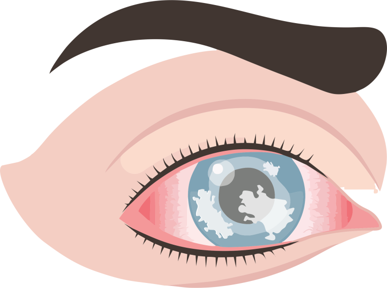
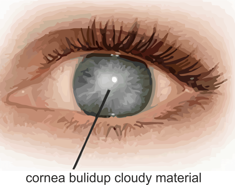
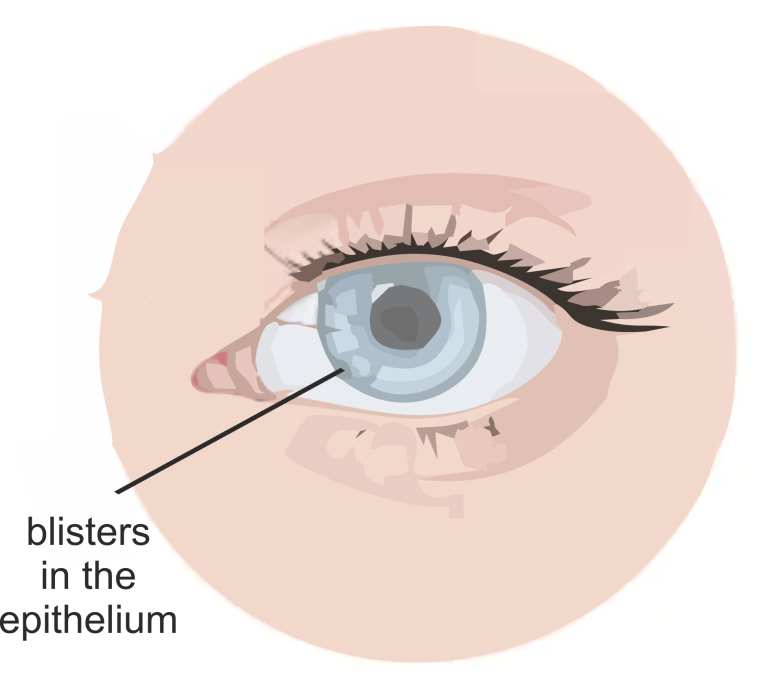
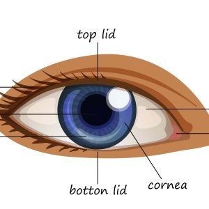
Keratitis is the inflammation of the cornea, the transparent front part of the eye covering the pupil and iris. It can be caused by various factors, leading to pain, blurred vision, and discomfort.

1. Infections: Bacterial, Viral, or fungal infections.
2. Injury: Scratches, cuts, or foreign objects in the eye.
3. Contact Lens Wear: Prolonged use or improper hygiene.
4. Dry Eyes: Insufficient tear production.
5. Underlying Conditions: Autoimmune diseases, like rheumatoid arthritis.

1. Eye Pain: Often severe and may worsen with blinking.
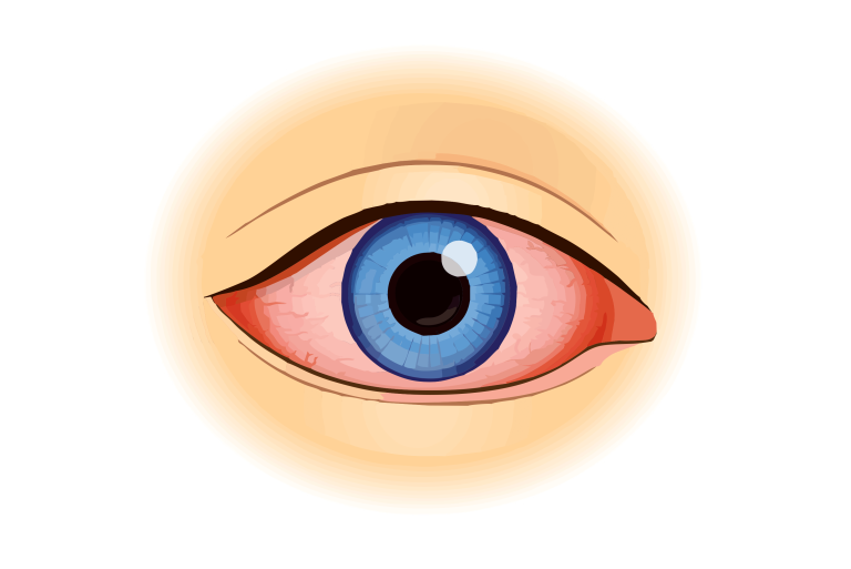
2. Redness: Bloodshot appearance of the eye.
3. Blurry Vision: Impaired or hazy vision.
4. Sensitivity to Light: Photophobia.
5. Excessive Tearing: Watery eyes.
6. Discharge: Pus or other discharge.

1. Bacterial Keratitis: Caused by bacterial infection.
2. Viral Keratitis: Caused by viruses like herpes simplex.
3. Fungal Keratitis: Caused by fungi, often in agricultural settings.
4. Acanthamoeba Keratitis: Rare, caused by amoeba found in water.

Keratoconus

Keratoconus eye-min

Keratoconus is a progressive eye disorder where the cornea, the clear front part of the eye, thins and bulges into a cone-like shape. This results in distorted vision and, if left untreated, can lead to significant visual impairment.

The exact cause of keratoconus is not fully understood, but several factors may contribute:

  1. Genetics: Family history of keratoconus increases the risk.
  2. Environmental Factors: Excessive eye rubbing, chronic eye irritation.
  3. Connective Tissue Disorders: Conditions affecting collagen.
  1. Blurred or Distorted Vision: Difficulty in seeing clearly.
  2. Increased Sensitivity to Light: Photophobia.
  3. Frequent Changes in Eyeglass Prescription: Rapid changes in vision.
  4. Ghosting of Images: Seeing multiple images.
  5. Eye Strain and Discomfort: Irritation or soreness.
  1. Corneal Topography: Mapping the curvature of the cornea.
  2. Slit-Lamp Examination: Detailed examination of the cornea’s structure.

Corneal Dystrophy

corneal-dystrophy-min

Corneal dystrophy refers to a group of genetic disorders that cause abnormal changes in the cornea, the clear front part of the eye. These changes can lead to visual impairment and may affect one or both eyes.

Corneal dystrophies are primarily caused by genetic mutations. These mutations lead to the accumulation of abnormal material in the cornea, disrupting its normal structure and function.

  1. Blurry or Hazy Vision: Loss of clarity in vision.
  2. Increased Sensitivity to Light: Photophobia.
  3. Corneal Clouding or Opacity: Gradual loss of transparency.
  4. Recurrent Corneal Erosions: Episodes of eye discomfort.
  5. Foreign Body Sensation: Feeling of a foreign object in the eye.
  1. Fuchs’ Endothelial Dystrophy: Affects the endothelial cells.
  2. Lattice Dystrophy: Deposits form in a lattice pattern.
  3. Map-Dot-Fingerprint Dystrophy: Abnormalities in the basement membrane.
  1. Corneal Examination: Via slit-lamp to assess changes.
  2. Genetic Testing: Identification of specific mutations.

Corneal Ulcer

corneal-ulcer-min

Corneal ulcers result from an infection or injury that causes damage to the cornea, the clear front part of the eye. Microbial agents like bacteria, viruses, and fungi are common culprits. Foreign objects, contact lens misuse, and dry eye syndrome can also contribute.

Corneal dystrophies are primarily caused by genetic mutations. These mutations lead to the accumulation of abnormal material in the cornea, disrupting its normal structure and function.

  1. Eye Pain: Sharp or constant pain in the affected eye.
  2. Redness: Increased redness and inflammation.
  3. Blurry Vision: Impaired vision due to corneal damage.
  4. Photophobia: Sensitivity to light.
  5. Excessive Tearing: Increased tear production.
  6.  
  1. Bacterial Corneal Ulcers: Caused by bacterial infection.
  2. Viral Corneal Ulcers: Result from viral infections like herpes.
  3. Fungal Corneal Ulcers: Caused by fungal infections.
  4. Acanthamoeba Keratitis: Caused by a waterborne amoeba.
  1. Slit-Lamp Examination: A magnified view of the cornea using a slit lamp to assess its shape, size, and any abnormalities.
  2. Confocal Microscopy: A non-invasive imaging technique to obtain detailed images of corneal layers, helping assess the extent of damage.
  3. Visual Acuity Test: Measures the clarity of vision to determine how much the corneal ulcer is affecting eyesight.
  4. Pachymetry: Measures the thickness of the cornea, helping in evaluating the severity of the ulcer.
  5. Schirmer’s Test: Measures tear production to evaluate dry eye syndrome, a potential contributing factor.


Fuchs' dystrophy

Fuchs’-Dystrophy

Fuchs’ Dystrophy is a genetic disorder affecting the corneal endothelium. The primary cause of Fuchs’ Dystrophy is a gradual loss of endothelial cells over time. 

1. Genetic Factors: Fuchs’ Dystrophy often has a familial tendency, indicating a genetic component. Specific genetic mutations may increase the risk of developing the disorder.

2. Age: Fuchs’ Dystrophy is more common in older individuals, and age is considered a significant risk factor. The condition tends to manifest later in life, typically around middle age or older.

3. Gender: Women are more commonly affected by Fuchs’ Dystrophy than men.

4. Inheritance: The disorder may be inherited in an autosomal dominant manner, meaning that a person has a 50% chance of inheriting the mutated gene from an affected parent.

5. Environmental Factors: While genetics play a crucial role, environmental factors may also contribute to the progression of the disease. Factors such as UV light exposure and environmental stressors could influence its development.

1. Indistinct or foggy vision, especially during the morning.
2. Sensitivity to light (photophobia).
3. Glare, especially in bright lighting conditions.
4. Difficulty seeing at night.
5. Eye discomfort or pain.

1. Early-onset Fuchs’ dystrophy: This type typically occurs in individuals aged 30 to 40 and progresses slowly over many years.

2. Late-onset Fuchs’ dystrophy: This type tends to develop in people over the age of 50 and may progress more rapidly.

1. Slit-Lamp Examination: A slit-lamp microscope allows the ophthalmologist to examine the cornea in detail. They will look for characteristic signs such as guttae (small bumps on the cornea) and evaluate the overall health of the cornea.

2. Corneal Endothelial Cell Count: This test measures the density of endothelial cells in the cornea. A decreased cell count is a hallmark of Fuchs’ Dystrophy.

3. Corneal Pachymetry: This test measures the thickness of the cornea. Thinning of the cornea is often seen in the advanced stages of Fuchs’ Dystrophy.

4. Specular Microscopy: This specialized imaging technique allows for detailed visualization of corneal endothelial cells. It helps assess the shape and health of these cells.

5. Acuity Test: A standard eye chart test is conducted to assess how well the patient can see at various distances. Fuchs’ Dystrophy can impact visual acuity, especially in the later stages.

6. Tonometry: This test measures intraocular pressure and helps rule out other eye conditions, such as glaucoma.

7. Topography: Corneal topography provides a detailed map of the cornea’s curvature, helping to identify irregularities.

Bullous Keratopathy

Bullous-Keratopathy-min

Bullous Keratopathy is a corneal disease characterized by permanent swelling of the cornea.

  1. Endothelial Damage: Damage to the endothelium impairs fluid regulation.
  2. Age-Related: Common in older individuals.
  3. Eye Surgery: Post-cataract surgery complication.
  4. Eye Trauma: Injury leading to corneal damage.
  1. Blurred Vision: Due to corneal swelling affecting light entry.
  2. Pain and Discomfort: Sensation of irritation or pain.
  3. Photophobia: Increased sensitivity to light.
  4. Redness: Inflammation of the eye.
  1. Corneal Pachymetry: Measures corneal thickness.
  2. Slit-lamp Examination: Assesses corneal health.
  3. Specular Microscopy: Evaluates endothelial cell density.
  4. Visual Acuity Test: Determines the extent of vision impairment.
Scroll to Top